|
|


|

Number of items in this site.
Cases:225
Images:1755
Cases:225
Images:1755
■Detailed data
| Materials |
Others |
| Microscopic findings |
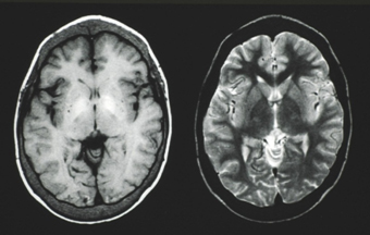
Can be seen in the basal ganglia iron deposition and thickening of the skull (head MRI scan).This case, the symptoms hemolytic microcytic hypochromic anemia HbF and HbA2 was found to increase and iron overload.Also periosteal reaction that radial thickening of the lid and skull X-rays in head-was also diagnosed with β-thalassemia. |

