|
|


|

Number of items in this site.
Cases:225
Images:1755
Cases:225
Images:1755
■Detailed data
| Materials |
peripheral blood(PB) |
| Stain |
May-Grünwald Giemsa (MG) |
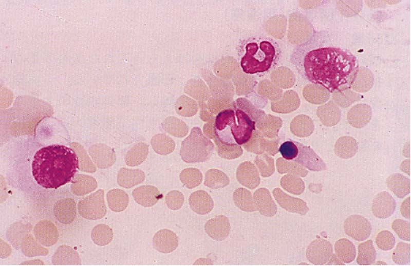
| Microscopic findings |
In the case of pancytopenia,
leukoerythroblastosis were observed. The cells in the 8-o’clock direction are slightly degenerated, but they are approximately twice the size of the blood neutrophils, the chromatin is coarse, and circular nucleoli can be seen in the stele. Based on the faint cytoplasm, the eccentric nuclei, the central circular nucleoli, and the like, we believe that these are derived from adenocarcinoma cells. The cells in the 1-o’clock direction are believed to be the same cells that have degenerated even further. They can be differentiated from reticular cells based on the roughness of the chromatin and the absence of vacuoles and phagocytic substances. 【MG.400×】 |

